

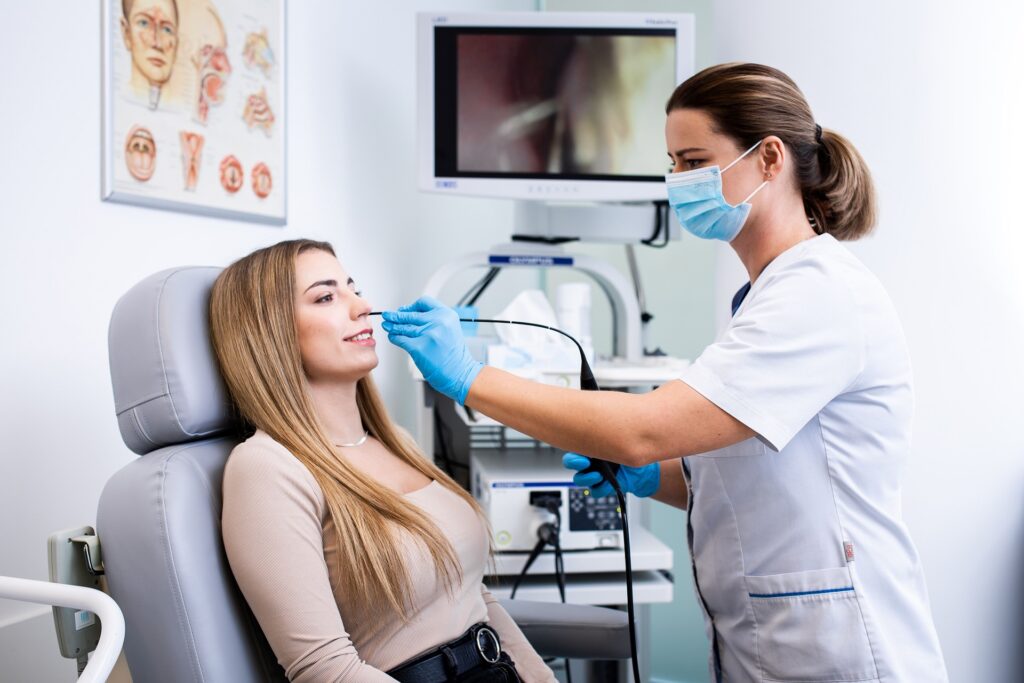


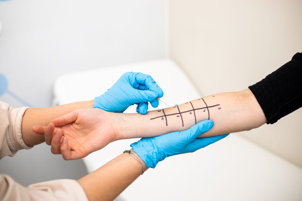


NBI is a safe for the patient, non-invasive diagnostic method using the properties of light waves, or more precisely – two of their lengths: 415 nm (blue) and 540 nm (green). The blood and tissues surrounding the capillaries absorb these colors in a different way than ordinary white light.
In this examination, during the observation of the mucosa with an endoscope, in addition to the usual white light, appropriate filters are additionally used and the tissue is illuminated in blue and green. The colored light penetrates into the mucosa by approximately 3 mm. Thanks to this, the structure and distribution of blood vessels (capillary and sub-epithelial networks) in the area under observation can be viewed in detail.
The colors show and enhance the image of the place where the blood supply is more intense than it should be. For the doctor, this sight is a warning signal. Too rich vascularity visible in a specific part of the tissue is a sign of something disturbing occuring in that place. In places where more blood vessels are formed, there is a high probability of cancer.
Cancer behaves like a parasite: to grow, it needs a supply of nutrients from its “host”. When there are abnormal cell divisions in the mucosa and the formation of a neoplastic lesion there, new, atypical blood vessels appear in the immediate area (they are usually less regular, more “looped” than healthy) – they provide an increased blood supply (nutriets for parasitic cancer cells).
The NBI technique uses this property of the tumor and shows precisely those areas of the mucosa that do not look normal and healthy. As a result, cancer can be found and treated quickly, even before major cancer changes start occurring.